
Introduction: Understanding the Condition
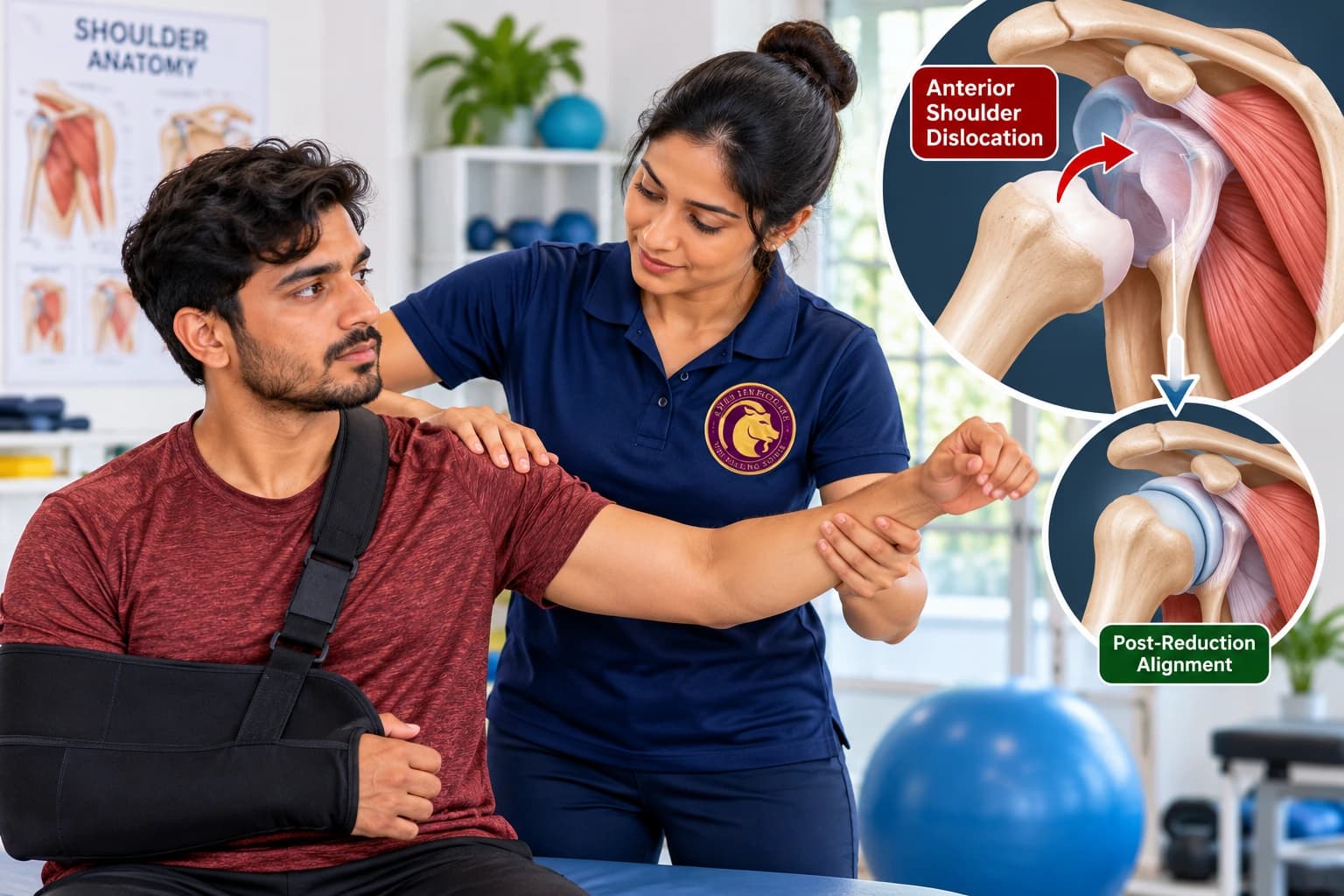
An acute glenohumeral dislocation is a high-energy traumatic orthopedic emergency representing the complete disarticulation of the humeral head from the glenoid cavity. Anterior dislocations account for over 95% of all cases, typically resulting from forced abduction, external rotation, and extension. Posterior dislocations, while less frequent, occur following seizures, electrical shock, or direct anterior impacts. Following prompt closed clinical reduction, structured multi-phase physical therapy is paramount. Inadequate or improper post-reduction management dramatically elevates the recurrence rate—surpassing 70% to 80% in active individuals under 25 years old. Rehabilitation focuses on phased mobilization, protection of injured static restraints, and progressive dynamic muscular reinforcement.
Common Symptoms
Key clinical indicators and functional presentations:
- Immediate, excruciating pain accompanied by total loss of active upper extremity function
- Prominent, visible asymmetry: Flattened lateral deltoid contour (squared-off shoulder) with a hollow space beneath the acromion
- Palpable anterior-inferior displacement of the humeral head into the subcoracoid region
- Compensatory posture: Patient supports the injured forearm in slight abduction with the opposite hand
- Sensory deficits or paresthesias over the lateral deltoid patch due to transient axillary nerve traction
- Post-reduction dull aching, joint stiffness, and acute muscular spasm throughout the shoulder girdle
Causes & Clinical Diagnosis
Etiological drivers and diagnostic assessment:
- High-energy contact sports collisions (rugby, football, wrestling) or falls onto an outstretched hand
- Sudden traction or violent abduction-external rotation loading
- Seizure activity or high-voltage electrical injuries predisposing to posterior dislocation
- Clinical diagnosis is established via neurovascular examination (checking axillary nerve sensation and radial pulse), confirmed by pre- and post-reduction orthogonal radiographs (AP, scapular Y, and axillary views) to identify associated fractures (Hill-Sachs and bony Bankart lesions).
Pathophysiology & Biomechanical Impact
During an anterior dislocation, the humeral head tears through the anterior-inferior labrum and capsuloligamentous complex, causing a classic Bankart lesion. Simultaneously, as the humeral head levers over the anterior glenoid rim, an impaction compression fracture occurs on the posterolateral aspect of the humeral head—termed a Hill-Sachs lesion. The axillary nerve, curving tightly around the surgical neck of the humerus, experiences significant mechanical elongation strain. The surrounding rotator cuff musculature undergoes acute reflex spasm followed by profound arthrogenic inhibition.
Evidence-Based Physiotherapy Protocols
At Aries PhysioCare, our physical therapists implement clinician-administered rehabilitation protocols:
- Phase I (Acute Protection & Healing): Therapist-managed sling immobilization protocols, monitoring axillary nerve recovery, and gentle active-assisted elbow/wrist/hand mobility
- Gentle submaximal isometric contractions of the rotator cuff and deltoid within neutral alignment to stimulate mechanoreceptors without displacing the joint
- Phase II (Controlled Range Restoration): Clinician-supervised passive and active-assisted range of motion strictly restricted to safe zones (avoiding external rotation past neutral and abduction past 90 degrees)
- Class-IV Laser Therapy and IFT applied to the anterior capsular region to control post-reduction hematoma and accelerate capsular fibroblastic bridging
- Phase III (Dynamic Neuromuscular Recalibration): Closed kinetic chain proprioceptive drills on stable surfaces to restore concave-compression force couples
- Scapular stabilization retraining focusing on the serratus anterior and lower trapezius to maintain an optimal glenoid platform beneath the humeral head
Home Care & Ergonomic Strategies
Supporting recovery through evidence-based ergonomic habits and self-care:
- Adhere strictly to prescribed sling wear times; do not remove the sling prematurely during sleep or community ambulation
- Perform gentle active wrist, hand, and finger movements several times daily to promote venous return and prevent secondary distal edema
- Never position the injured arm in abduction combined with external rotation (the throwing position)
- Apply cryotherapy packs over the anterior shoulder for 15 minutes three times daily to minimize residual swelling and joint effusions
- Sleep strictly on your back with the immobilized arm supported on a pillow to prevent nocturnal posterior sagging or roll-over trauma
- Avoid pushing, pulling, lifting, or reaching for objects with the injured arm until cleared by your physical therapist
When to Contact Us
Recovering from a recent shoulder dislocation? Contact Aries PhysioCare today for a structured, evidence-based post-reduction rehabilitation protocol designed to prevent recurrent instability.