Introduction: Understanding the Condition
Frozen shoulder, clinically known as adhesive capsulitis, is an insidious, disabling condition characterized by progressive fibrosis, thickening, and contracture of the glenohumeral joint capsule. Affecting approximately 2% to 5% of the general population—with a marked predilection for individuals with diabetes mellitus or thyroid dysregulation—it severely restricts both active and passive shoulder mobility. Normal glenohumeral joint volume of 15 to 30 milliliters shrinks dramatically to less than 5 to 10 milliliters as the axillary fold adheres and obliterates the inferior recess. Clinical management requires specialized, stage-matched physiotherapy to preserve neurovascular health, mitigate chronic pain, and systematically restore capsular compliance without inducing inflammatory flare-ups.
Common Symptoms
Key clinical indicators and functional presentations:
- Severe, unrelenting deep shoulder ache that intensifies notably at night, disrupting restorative sleep
- Marked global restriction of both active and passive glenohumeral motion, especially external rotation, abduction, and internal rotation
- Inability to perform functional daily tasks such as reaching behind the back, fastening clothing, or grooming hair
- Compensatory shoulder hiking and excessive scapulothoracic elevation during attempted arm elevation
- Radiation of dull, aching pain down the lateral deltoid insertion toward the mid-upper arm
- Progression through distinct clinical phases: painful freezing phase, stiff frozen phase, and gradual thawing phase
Causes & Clinical Diagnosis
Etiological drivers and diagnostic assessment:
- Idiopathic primary capsular inflammation involving autoimmune and fibroblastic dysregulation
- Secondary systemic risk factors including Type 1 and Type 2 diabetes mellitus, Hashimoto's thyroiditis, and Dupuytren's contracture
- Secondary intrinsic or extrinsic factors such as prolonged shoulder immobilization following breast surgery, cardiac procedures, or distal upper-limb fractures
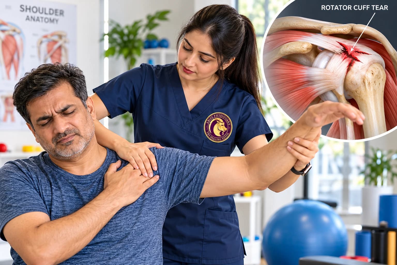
- Clinical diagnosis involves passive range of motion assessment confirming the capsular pattern (external rotation most limited, followed by abduction and internal rotation), standard orthopedic provocative testing, and MRI evaluation to rule out rotator cuff tears while confirming axillary pouch thickening.
Pathophysiology & Biomechanical Impact
Adhesive capsulitis is primarily a fibroproliferative condition driven by an imbalance of pro-inflammatory cytokines, transforming growth factor-beta (TGF-beta), and matrix metalloproteinases. Chronic low-grade synovitis triggers extensive fibroblast and myofibroblast proliferation within the anterior-superior capsule and coracohumeral ligament. The resulting deposition of dense type I and type III collagen bundles obliterates the normal capsular elasticity, creating rigid contractures across the axillary pouch and rotator interval. This capsular shortening dramatically restricts humeral head gliding, driving excessive superior humeral migration and secondary subacromial friction during attempted arm elevation.
Evidence-Based Physiotherapy Protocols
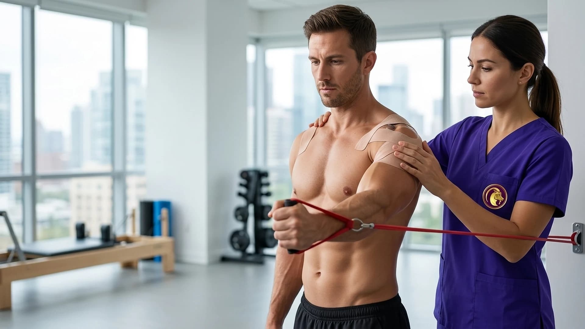
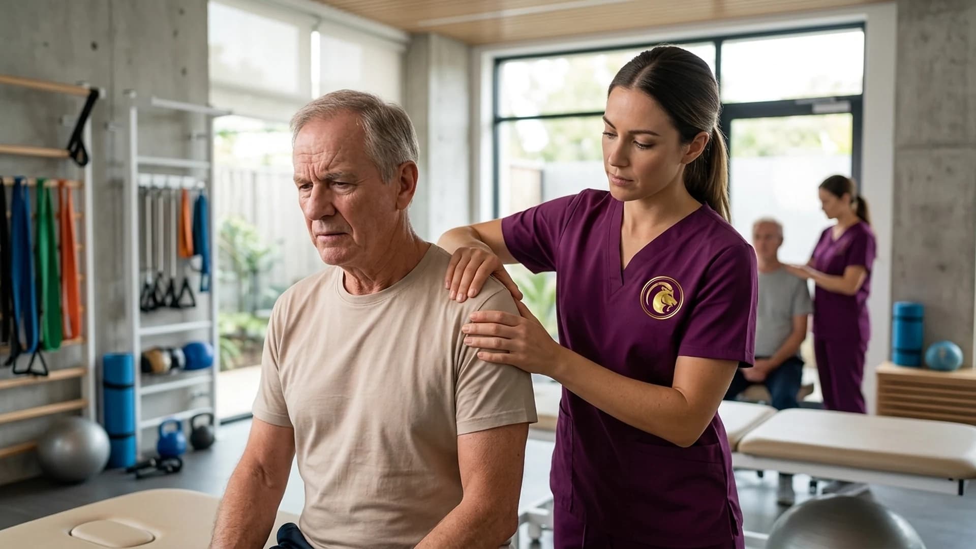
At Aries PhysioCare, our physical therapists implement clinician-administered rehabilitation protocols:
- Maitland manual therapy: Application of gentle Grade I and Grade II oscillatory glenohumeral joint mobilizations to desensitize capsular mechanoreceptors during the acute freezing phase
- End-range Grade III and Grade IV inferior and posterior capsular glides (Kaltenborn techniques) administered during the chronic frozen and thawing stages to elongate dense collagenous adhesions
- Mulligan Mobilization with Movement (MWM) to restore pain-free physiologic humeral elevation and external rotation through therapist-assisted glide corrections
- Targeted Radiofrequency Therapy (Tecar) and Class-IV Deep Tissue Laser Therapy to enhance microvascular perfusion and increase capsular collagen extensibility prior to manual mobilization
- Calibrated Interferential Therapy (IFT) and high-voltage pulsed galvanic stimulation for non-pharmacological pain inhibition and paraspinal muscle relaxation
- Progressive scapulothoracic neuromuscular re-education under direct clinician supervision to re-establish normal 2:1 scapulohumeral rhythm and eliminate compensatory shoulder hiking
Home Care & Ergonomic Strategies
Supporting recovery through evidence-based ergonomic habits and self-care:
- Position the affected arm on a supportive contour pillow during sleep to maintain neutral humeral abduction and prevent painful nocturnal compression
- Apply moist heat packs over the anterior and lateral shoulder girdle for 15 to 20 minutes prior to physical therapy sessions to soften periarticular connective tissue
- Utilize localized cryotherapy applications for 10 to 15 minutes post-activity if reactive inflammatory discomfort occurs
- Optimize desk and computer workstations with adjustable armrests that support the forearm at a 90-degree elbow flexion angle without shoulder elevation
- Avoid carrying heavy loads, briefcases, or grocery bags with the affected arm to prevent sudden traction on contracted capsular tissues
- Strictly avoid unsupervised ballistic stretching or aggressive arm pulling, which damages contracted capsular fibers and provokes secondary inflammatory cascades
When to Contact Us
Do not allow shoulder stiffness to progress into permanent mobility loss. Contact Aries PhysioCare today to schedule a comprehensive clinical shoulder assessment with our dedicated orthopedic physical therapists.